

Short answer
The effect of a visual impairment on the brain depends on the type and cause of the visual impairment. Visual impairments caused by brain damage are difficult to treat. A visual prosthesis, an implant that directly stimulates brain areas through electrical signals, may offer hope for blind individuals to perceive shapes and letters again.
Longer answer
A visual impairment means that someone can see only partially or not at all. When a person can see less than 10% of what we normally perceive, the World Health Organization (WHO) classifies this as blindness. Blindness is a significant disability that, unfortunately, around 43 million people worldwide still have to live with. Although visual impairments all involve a loss of sight, the underlying cause is often not the same.
An anatomical over-‘view’
To better understand visual impairments, we need a better over-‘view’ of our brain (see also Figure 1)! The first brain region involved in seeing is our eyes. That’s right — part of the eyes is already a part of the brain. The retina at the back of our eyes is the first link in a chain of brain regions that process light from the outside world. The retina contains neurons (brain cells) that perform computations and are directly connected to the rest of our brain. Virtually all visual information then travels through the optic nerve, crosses at the optic chiasm, and arrives at the next station: the thalamus. Within the thalamus lies a small brain nucleus called the Lateral Geniculate Nucleus (LGN), which consists of multiple layers of brain cells. These layers process different elements of the visual information. The final destination of visual information is the large, folded outer layer of the brain that we call the cortex — or cerebral cortex. The cortex is the place where we perceive and interpret visual information, in a process we still do not fully understand.
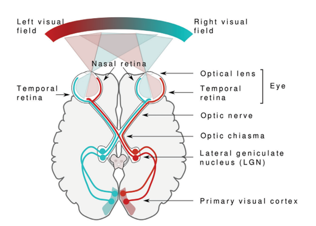
Figure 1: Brain Anatomy of Vision.
Light from the outside world is projected through the optical lens onto the retina at the back of both eyes. Visual information then travels via the optic nerve, through the optic chiasm, to the LGN in the thalamus. From the LGN, this information is sent to the visual cortex (primary visual cortex). Visual information from both eyes converges at the optic chiasm, where it is separated based on the visual field. Visual information from our right visual field (in blue) is processed in the left hemisphere of the brain after the optic chiasm, and vice versa. This separation of information continues in the thalamus and cortex. In addition, visual information enters the brain upside down from our eyes. Our brain corrects for this. This image is taken from the BrainFacts book (2018) from the Society for Neuroscience.
From glasses to brain implants
In the entire process that leads to “seeing,” many things can go wrong. In some visual impairments, problems occur even before light reaches the retina. For example, abnormalities in the eyeball or lens can make it difficult to see things at a distance (nearsightedness) or up close (farsightedness). These types of impairments are now easily treatable in the Netherlands with a quick visit to the optometrist: a good pair of prescription glasses, contact lenses, or even laser surgery can often fix the problem in the blink of an eye!
The situation is more complex when the visual impairment is caused by something going wrong in one of the brain’s own processing steps. For instance, the retina can be damaged by macular degeneration. The macula (or yellow spot) is the part of the retina responsible for sharp central vision. Damage can also occur in the retinal blood vessels due to diabetes (also known as diabetic retinopathy). Another common cause of blindness is glaucoma — an eye disease that damages the optic nerve. In all these cases, parts of the brain’s visual system are affected and (at least for now) they cannot be treated easily.
Still, even for this group of blind individuals, there is a glimmer of hope. At the Netherlands Institute for Neuroscience, we are working on designing a visual prosthesis. A prosthesis is an artificial aid that replaces or supports a body part — like dentures, a hearing aid, or a prosthetic leg. A visual prosthesis is an implant that is surgically placed directly into the brain to compensate for the lack of visual input. Brain cells communicate with each other through electrical signals (you can read more about that here). With this implant, we deliver tiny electrical pulses to a specific part of the brain to mimic the missing visual signals. You can imagine that such an implant can be placed at different points along the stream of brain areas that process visual information (Figure 1). Among the most successful types of visual prosthesis are retinal implants and optic nerve implants for example.
The Visual Prosthesis
Within Pieter Roelfsema’s research group, we are working on a cortical visual prosthesis that we place all the way at the back of the brain, in the primary visual cortex (for a review of the different the steps in our visual system, see Figure 1). Through many years of research in animals and later in humans, we’ve learned that the primary visual cortex contains a sort of map of the outside world. Brain cells in this area respond very precisely to visual stimuli at specific locations in our visual field. For example, there are brain cells that respond when you see a light spot in the upper left of your visual field, and others that respond when a spot appears in the lower right. This map comes in handy! It means we can deliver electrical pulses to cells that each correspond to a different part of the visual field. Research has shown that when we stimulate these cells, people perceive a light spot at that exact location in their visual field! By combining these light spots in a meaningful way, we can simulate a form of vision.
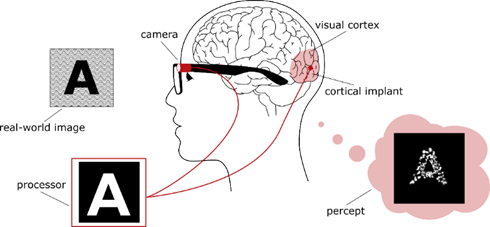
Figure 2: A Visual Prosthesis
Images from the outside world (in this case, the letter ‘A’) are captured by a camera mounted on a pair of glasses. This camera converts the signals into a pattern that can be used to activate brain cells through small electrical pulses. The pattern of pulses then allows blind individuals to perceive the letter ‘A’.
Video 2: Eye Movements of a Monkey with a Visual Prosthesis
The cells in the monkey’s visual cortex can be stimulated in such a way that the monkey “sees” different letters (the monkey does not see the green dots on the screen—each green dot represents one electrode). The monkey indicates which letter it “sees” by making an eye movement toward that letter and receives a reward each time it gets it right.
To understand the visual cortex and to develop a visual prosthesis, monkeys are often used. Their visual system (as shown in Figure 1) closely resembles that of humans, and the animals can be trained to indicate what they see through hand or eye movements. This research has led to the first successful brain implants being placed in blind people! For instance, the Spanish woman Bernardeta Gomez was the first person to receive such an implant, and she reported that—just like the monkeys—she was able to recognize patterns and letters. She had been completely blind for 16 years, but with the implant, she was even able to play a simplified version of the computer game Pac-Man.
In follow-up research at the Netherlands Institute for Neuroscience, we are working on improving this type of visual prosthesis. We do this by optimizing both the hardware and the software. For example, we are testing different materials for the implants so they can generate better images, last longer, and be more compatible with the brain. We are also improving the algorithms that convert images from the outside world into patterns that can be more easily interpreted by the brain.
Video 2: Improving a Visual Prosthesis
In a collaboration between the Netherlands Institute for Neuroscience in Amsterdam and the Donders Institute in Nijmegen, we are optimizing how images from the outside world can best be used by a prosthesis. For example, the visual prosthesis converts video footage of this swimming penguin into images that can be projected onto the brain. The video on the right shows what someone with a visual prosthesis will eventually be able to see.
A Glimpse into the Future
Although we are still far from having a complete picture, we believe this research sheds a promising light on the future for people with severe visual impairments. The effect of a visual impairment on the brain—and how we can treat it—depends greatly on the type and cause of the impairment. It all hinges on where in the eyes and/or brain something goes wrong within the many links that make vision possible (the eyeball, the retina, the thalamus, and the cerebral cortex). For people who have lost their sight due to damage to the eyes, a visual prosthesis offers hope—the hope of being able to perceive shapes and letters once again!
Read more?
(Dutch) https://hersenvrienden.nl/actueel/hoe-werkt-de-visuele-prothese/
(Dutch) https://www.medischcontact.nl/actueel/laatste-nieuws/artikel/blind-zijn-en-toch-zien
(English) https://elifesciences.org/articles/85812